
(727) 514-0100
A nuclear stress test, also called myocardial perfusion imaging, allows doctors to see images of your heart while it is at rest and after stress or exercise. The test can show how well your heart is pumping blood, whether there is damaged heart muscle, and whether blood flow to the heart may be reduced due to narrowed or blocked arteries.
Your doctor may recommend a nuclear stress test to better understand how your heart is functioning, especially if you are experiencing symptoms such as chest pain or shortness of breath.
This test may help diagnose or evaluate:
As with most medical procedures, some risks are possible. However, complications from a nuclear stress test are rare.
Your care team may ask questions about your medical history and exercise habits to determine the appropriate level of stress for the test. They may also listen to your heart and lungs before the test begins.
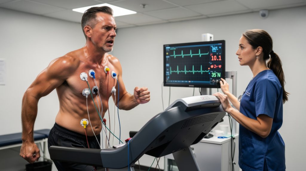
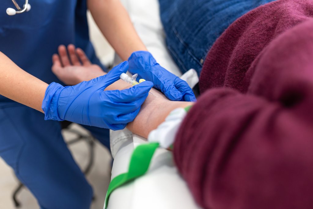
A technician will place an IV line in your arm. Electrodes will be placed on your chest to monitor your heart’s electrical activity, and a blood pressure cuff will be used to monitor your blood pressure throughout the test. In some cases, small areas of the chest may need to be shaved so the electrodes can stick properly.
A radiotracer will be injected before the first set of resting images. The injection may feel cold at first. After about 20 to 40 minutes, imaging will begin.
The first set of images will be taken while your heart is at rest. After that, your heart will be stressed either through exercise or medication.
You may walk on a treadmill or ride a stationary bike. The intensity will gradually increase until you reach your target heart rate. Another tracer will then be injected, and you will continue exercising briefly before the stress images are taken.
If you are unable to exercise, medication may be used to mimic the effects of exercise by increasing blood flow to your heart. Some patients may experience temporary symptoms such as a headache or shortness of breath. A second set of images will be taken after the medication has taken effect.
After the test, you can usually return to normal activities unless your doctor gives you different instructions. The tracer naturally leaves your body through urine or stool. Drinking water can help flush it from your system.
During the imaging portion of the test, you will sit upright while images of your heart are taken. If possible, your arms will be positioned above your shoulders on a cushioned armrest. It is important to remain still during imaging, as movement can affect the quality of the images.
Once both the rest and stress images are completed and processed, your test will be finished.
To learn more or get started simply fill out the short form below.